
Yeeran
Application of YEERAN ULTIMUS 9 LAB small animal super-resolution ultrasound imaging system in microvessels such as brain, heart, kidneys, and tumors
Release Time:
2024-12-10 14:40

"In the exploration of life sciences, the complexity of the microcirculatory system often becomes a key difficulty in research."
The ULTIMUS 9 LAB small animal super-resolution ultrasound imaging system launched by YEERAN has once again achieved important technological application progress in 2024, opening up a new direction for research in the field of life sciences. Since its launch last year , with its innovative URM (ultra-resolution microscopy) technology, the ULTIMUS 9 LAB has achieved an imaging accuracy of ≤10 microns, clearly observing and analyzing microvessels, and revealing the complex structure of the microvascular system. This technological advancement has opened up a new perspective for the study of microvascular-related diseases such as neurological, cardiovascular, tumor, and kidney diseases, and promoted scientific research progress in related fields.
As a model of whole-body ultrasound application, the MUSE soft beam synthesis platform built into the ULTIMUS 9 LAB can intelligently adapt to various application scenarios with its excellent computing power and multi-level optimization processing algorithm, providing researchers with the best imaging solution. The addition of this platform has made a qualitative leap in image quality and high-end functions, making every experiment more accurate and efficient.
ULTIMUS 9 LAB has demonstrated its excellent performance in multiple research fields. For example, in the study of carotid thrombosis, the system successfully captured Doppler images before and after thrombus formation, providing valuable data for understanding the dynamic process of thrombus formation. In terms of mouse heart imaging, both normal hearts and myocardial infarction models have demonstrated its excellent ability in microstructure and function assessment. In addition, the system also performs well in imaging mouse kidneys, cerebellum, stroke, colorectal cancer, and muscle tissue, providing more detailed and clearer images than traditional ultrasound, helping researchers to explore disease mechanisms and development processes more deeply.
The following is a demonstration of technical applications:
01 Multi-port Doppler imaging of Carotid artery thrombosis (before and after )
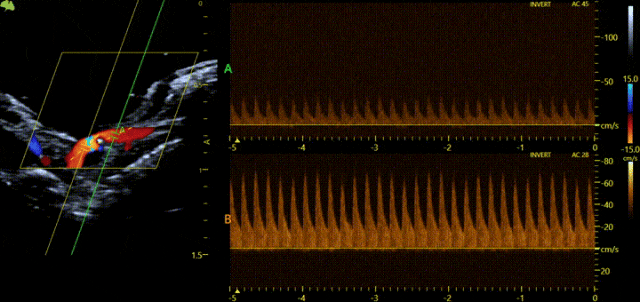
Ferric chloride (FeCl3) has been widely used to make thrombosis models, especially in scientific research. This model usually induces thrombosis by applying a certain concentration of ferric chloride solution on the surface of the blood vessels of animals (such as mice, rats, rabbits, etc.). The process of thrombosis is often analyzed by real-time ultrasound video recording and blood flow images, especially the changes at different time points. However, traditional ultrasound can only measure the blood flow spectrum before and after thrombosis at different time periods. Sometimes the heart rate is different at different time periods, and the model itself has the situation of vascular thrombosis recanalization. It is very likely that the observation time is missed when the sampling volume is moved. Our latest multi-gate Doppler technology can obtain the Doppler spectrum of up to four sampling points in real time in the same cardiac cycle, providing users with more hemodynamic diagnostic information. At the same time, we also have PW/TD dual synchronization, which can simultaneously evaluate ventricular wall motion and hemodynamics, and can measure LV diastolic dysfunction faster and more accurately.
02 Mouse heart
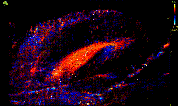
Normal mouse heart super-resolution ▲
Super-resolution of the heart in infarcted mice▼
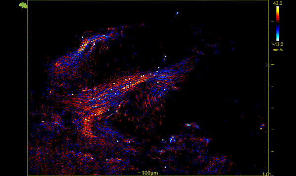
Ultrasound can be used to determine the health of the heart muscle by tracking the flow of gas-filled microbubbles through the small blood vessels that perfuse the heart muscle with oxygenated blood. The method used in this study is based on ultrasound resolution microscopy (URM), a super-resolution imaging technique that uses micron-sized microbubbles (MBs) to surpass the resolution limit of traditional ultrasound imaging and achieve a spatial resolution of hundreds of microns in a moving heart. This resolution can distinguish blood vessels hundreds of microns apart in a beating heart. This study provides an exciting attempt at the future of myocardial imaging.
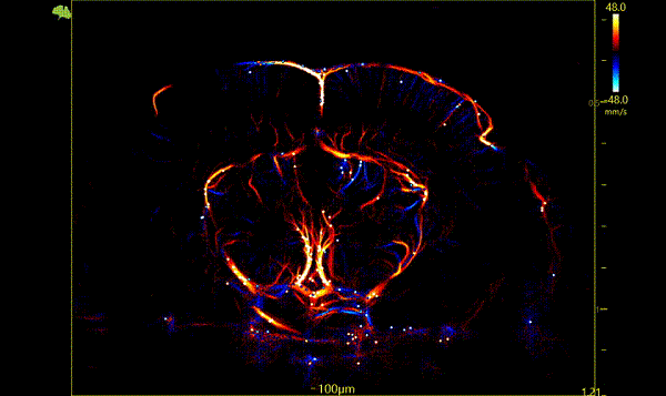
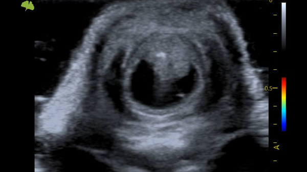
03 Mouse kidney
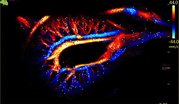
Ultra-resolution of kidney in cross section
Traditional renal function testing methods are insufficiently sensitive to early microcirculatory changes and renal fibrosis, and these changes are key factors in the progression of CKD, including microvascular sparseness and tissue fibrosis. These pathological changes often lead to a significant decline in renal function. It's happened before. This study provides a new, non-invasive ultrasound imaging technology that can early diagnose and monitor microcirculatory changes in CKD. Through ultra-resolution ultrasound imaging (URM), the microvascular structure of the kidney can be observed in unprecedented detail. The above research results demonstrate the potential of advanced ultrasound technology in the early diagnosis and management of CKD, especially in improving resolution and microvascular assessment. importance.
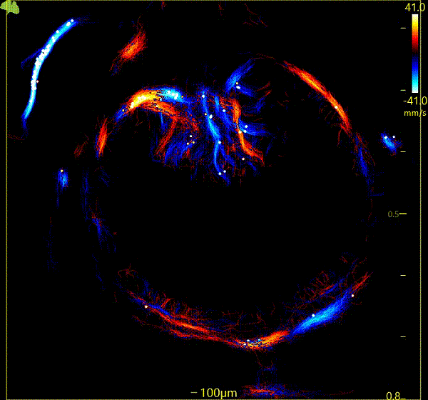
04 Mouse cerebral blood flow imaging
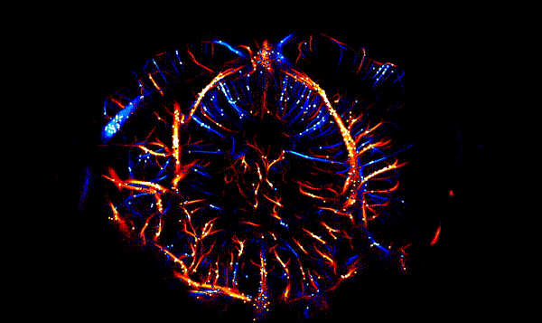
Super-resolution of normal mouse brain
Since the brain is an organ with high metabolic demands, maintaining normal cerebral blood flow is essential for its function. URM can provide wide spatial coverage of brain regions and high spatial resolution, thereby accurately monitoring and mapping cerebral blood flow in various brain regions.
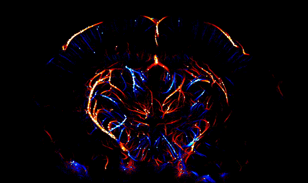
Super-resolution image of normal mouse brain ▲
super-resolution image of left cerebral stroke ▼
Traditional laser speckle can only identify the microvessels on the surface of the brain, and we have no idea about the blood flow in the deeper layers. This has also led many researchers to use laser speckle only to detect the surface cerebral blood vessels when making stroke models, resulting in misjudgment of the results. When the final sample was taken, it was discovered that the model making was not completely successful. URM can be used for early detection and diagnosis of stroke, and by identifying and quantifying damaged microvascular blood flow, it can help assess the severity and recovery of stroke.
05 Mouse rectal cancer
2D image of rectal cancer longitudinal section▲
Super-resolution image of rectal cancer longitudinal section▼

2D image of rectal cancer cross section▲
Super-resolution image of rectal cancer cross section▼
Angiogenesis is a very important early physiological marker of tumors. URM can detect and quantify changes in microvascular blood flow in tumors with high resolution, thereby helping experimenters to detect and diagnose tumors early or predict the area where tumors occur. At the same time, URM can be used to monitor the impact of cancer treatment on tumor microvessels, evaluate treatment effects, and adjust research plans in a timely manner. By regularly imaging (non-invasive, non-destructive) microvascular changes in tumors, a complete chain of experimental evidence can be provided for tumor quality. By imaging the microvascular structure and blood flow pattern in detail in tumors, researchers can have a deeper understanding of the generation and classification of tumors.
06 Mouse thigh

Ultra-resolution of the thigh in longitudinal section

Ultra-resolution of the thigh in cross-section
In previous ultrasound applications, due to resolution reasons, people can only evaluate blood flow changes in the femoral artery and femoral vein. For some vascular diseases, such as diabetic foot complications caused by diabetes, the affected limbs suffer from insufficient arterial perfusion due to large blood vessels and microvascular lesions in the lower limbs, resulting in foot infection, ulcers or deep damage. URM breaks the diffraction limit of traditional ultrasound and presents microvessels to people from another new perspective, allowing researchers to have a more intuitive understanding of diabetic foot caused by diabetes.
The breakthrough micron-level imaging of ULTIMUS 9 LAB brings more possibilities to life science research. YEERAN will continue to be committed to innovation and breakthroughs, using ULTIMUS 9 LAB as a platform to explore the infinite possibilities of life science with researchers around the world.
Provider of overall solution


Contact Us
ADD:Room 1208, 12th Floor, Building 3, World Overseas Chinese Business Center, Tongzhou District, Beijing, China
Postal Code:101100
Tel :+86-010-69549099
Copyright © Beijing Yeeran Technology Co., Ltd. Powered by 300.cn SEO Business license